

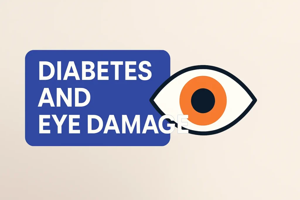
How Diabetes Damages Your Eyes: Beginner’s Guide to Diabetic Retinopathy — 7 Scary Warning Signs I Ignored Before My Eye Exam
If you’re living with diabetes, chances are you spend more time thinking about A1C levels and counting carbs than worrying about the tiny blood vessels hiding behind your eyeballs. I know I did. For years, I chalked up blurry vision to bad lighting, cheap screens, or just “being tired.” Squinting at restaurant menus? I blamed the font. Turns out, my eyes were waving red flags the whole time—I just wasn’t paying attention.
It wasn’t until I finally dragged myself to a dilated eye exam (after months of putting it off, of course) that I got the wake-up call: I had diabetic retinopathy. Damage had been quietly building up like that weird smell in your fridge you keep meaning to check out but never do—until it’s too late.
In this guide, I’ll walk you through what diabetic retinopathy really is (no sugar-coating it—pun intended), the 7 warning signs I ignored like a champ, what can happen if you keep kicking the can down the road, and—most importantly—how you can start protecting your vision today. I know you’re busy. I know this stuff is scary. So I promise to keep it clear, real, and doable—in the time it takes to finish your coffee.
Table of Contents
What Exactly Is Diabetic Retinopathy?
Diabetic retinopathy is what happens when long-term high blood sugar damages the tiny blood vessels in the retina, the light-sensitive layer at the back of your eye. Think of the retina as the camera sensor inside your eye. When its wiring and plumbing are stressed, the picture gets noisy, distorted, or goes dark in patches.
Doctors often divide diabetic retinopathy into two big buckets: non-proliferative (early) and proliferative (advanced). In the early phase, blood vessels weaken and leak small amounts of fluid or blood. In the advanced phase, new fragile vessels grow where they shouldn’t, creating scar tissue and a very real risk of sudden vision loss.
Many people never feel pain. That’s the cruel part. Your eyes can look “normal” in the mirror while tiny changes accumulate at the back of the eye. A lot of us only take it seriously when something dramatic happens: a big floater, a dark curtain in our vision, or a scary line from the eye doctor like, “I’m concerned about bleeding at the back of your eye.”
In clinic stories, the pattern repeats: 10–15 years of diabetes, eyesight “good enough” for daily life, and then a single exam where the photos tell a different story. If you have diabetes, this condition is common enough that it deserves a firm spot on your annual to-do list, right alongside lab work and medication refills.
“Your vision can feel fine while damage quietly stacks up in the background.”
- You can’t judge your retina’s health by how clearly you see today.
- Early stages are treatable; late stages are harder and more expensive.
- Annual dilated eye exams are non-negotiable if you live with diabetes.
Apply in 60 seconds: Open your calendar app and block one hour this month labeled “Dilated eye exam (diabetes).”
How Diabetes Silently Damages Your Eyes
So how does blood sugar in your bloodstream cause trouble inside your eyes?
Over time, high glucose acts like sticky syrup on the inside of tiny blood vessels. The vessel walls stiffen and weaken. Little outpouchings called microaneurysms form. Some vessels start to leak fluid or blood. Others close off entirely, starving parts of the retina of oxygen.
Imagine a neighborhood where half the streets slowly crumble and the rest get random potholes. Delivery trucks still try to bring packages, but routes get longer and less reliable. In your eye, this “bad infrastructure” means light signals don’t travel cleanly from the retina to your brain. You experience that as blur, distortion, or missing pieces of the image.
When the retina senses it’s not getting enough oxygen, it panics and releases signals that tell your eye to grow new blood vessels. That might sound helpful, but these new vessels are fragile. They bleed easily, tug on the retina, and can cause scar tissue that literally pulls the retina off the back wall of the eye.
The wild part? This can be happening while you’re still driving, reading, and scrolling on your phone without much trouble. I remember thinking, “I can read the menu, so my eyes are fine,” while my retina was quietly collecting tiny leaks like a ceiling with slow water damage.
Show me the nerdy details
At the microscopic level, chronic high blood sugar damages endothelial cells lining retinal capillaries and alters the blood-retina barrier. Pericytes, the “support cells” wrapped around capillaries, are particularly sensitive. Their loss contributes to microaneurysm formation and capillary dropout. The resulting ischemia drives up levels of growth factors such as VEGF, which in turn promotes abnormal new vessel formation and increased vascular permeability. Modern treatments like anti-VEGF injections work by blocking those growth signals.
- Leaky vessels and blocked vessels both harm the retina.
- Your eye tries to fix the problem by growing fragile new vessels.
- Modern treatments target those abnormal growth signals.
Apply in 60 seconds: Write down your last A1C and blood pressure readings; bring them to your next eye visit so your doctor can see the full picture.
7 Scary Warning Signs I Ignored Before My Eye Exam
Here are the seven red flags I brushed off, all easy to blame on “age,” “screens,” or “a bad night’s sleep.” If any of these feel familiar, your eyes deserve a closer look.
- Blur that comes and goes during the day. Some mornings, my phone text looked fuzzy, then sharpened by lunch. I blamed coffee. In reality, shifting blood sugar can temporarily change the shape of the lens inside your eye.
- Needing more light than friends the same age. In dim restaurants, everyone else was fine with candles and soft light. I was the one asking the server to “crank the lights a bit” or turning on my phone flashlight over the menu.
- Dark smudges and floaters that didn’t quite go away. I remember a tiny thread-like floater that drifted across my vision while I typed. I blinked it away. Weeks later, it was back, joined by a small cloudy patch, like a thumbprint on my glasses.
- Lines that looked slightly wavy on the page. When I lined up spreadsheets, some rows looked like they dipped or bowed. I convinced myself it was the monitor. Distortion like this can be a sign of swelling in the central retina.
- Colors losing their “pop.” Reds felt more like brick than cherry. I figured my screen profile was off. Subtle color changes can happen when the macula is under stress.
- One eye doing more work than the other. I started closing one eye when reading small labels because “it felt sharper.” That was my brain quietly favoring the eye with less damage.
- Headaches after close work, not just stress. I told myself they were laptop headaches. In reality, my eyes were straining to compensate for patchy vision.
None of these felt dramatic enough to cancel a workday or rush to the emergency room. That’s exactly why they’re dangerous. They are easy to normalize, especially if you’re the responsible type who always powers through discomfort.
If you recognize even one of these signs and you have diabetes, treat it as a friendly alarm bell, not a verdict. The whole point of catching them now is to keep you driving, reading, and recognizing your loved ones’ faces for decades.
“The scariest part wasn’t the diagnosis. It was realizing how long I’d been explaining away the clues.”
- Floaters, waves, and faded colors are worth a professional look.
- Symptoms may come and go, even while damage is steady.
- One quick exam can reset years of quiet anxiety.
Apply in 60 seconds: List any odd visual symptoms you’ve noticed in the last 3 months and bring that list to your eye appointment.
Coverage, Costs, and Why Waiting Gets Expensive
This part is where money and medicine collide. Many readers don’t delay eye care because they’re lazy; they delay because they’re scared of the bill. That’s reasonable. But with diabetic retinopathy, procrastination often makes care more expensive, not less.
In early stages, your main costs are routine: a dilated eye exam, perhaps retinal photos, maybe an optical coherence tomography scan (a detailed “cross-section” image of your retina). These are often covered as part of diabetes management benefits under medical insurance, not just vision plans. Your responsibility is usually the deductible and copay.
Once the disease advances, the price tag climbs quickly. Think of monthly injections into the eye, laser procedures, or even surgery to remove blood and scar tissue from inside the eye. Each of those comes with its own billing code, prior authorization dance, and potential out-of-pocket surprises.
I’ve seen people try to “save” a few hundred dollars by skipping exams, only to face multi-thousand-dollar treatment plans later. It’s like skipping oil changes to avoid a small fee, then paying for an engine replacement. Except in this case, the engine is your vision.
Good news: insurers and government programs know diabetes affects the eyes. Many explicitly cover at least one comprehensive exam per year for people with diabetes. Some Medicare Advantage and commercial plans even highlight this in their preventive care benefits. The trick is to confirm your coverage tiers and fee schedule before you sit in the exam chair.
- Many plans treat diabetic eye exams as medical, not “optional” vision perks.
- Checking your coverage and deductible upfront reduces bill anxiety.
- Every year you delay increases the odds—and cost—of advanced treatment.
Apply in 60 seconds: Call the number on your insurance card and ask, “How many diabetic eye exams per year are covered, and what’s my estimated out-of-pocket cost?”
Money Block: 60-Second Eligibility Checklist for Covered Eye Exams
Before you start comparing eye clinic quotes, check whether you already qualify for a covered exam under your current plan. This is a quick, binary checklist you can run through while you wait for the microwave.
Eligibility Checklist
- ✔ You have a documented diagnosis of type 1 or type 2 diabetes.
- ✔ You are enrolled in a medical insurance plan, Medicare, or a national health system.
- ✔ Your plan includes outpatient specialist visits (often under “specialist copay”).
- ✔ Your primary care doctor or endocrinologist can provide a referral if required.
- ✔ You have not used up your annual limit for covered diabetic eye exams.
If you answered “yes” to at least three of these, you are very likely eligible for a covered comprehensive eye exam.
Apply in 60 seconds: Grab your insurance card and note three things: plan type, member services phone number, and whether referrals are required. Save this table and confirm the current rules on your provider’s official site or hotline.
Many older adults also want to understand how diabetic eye disease fits alongside other age-related issues like cataracts and macular degeneration. If you’re navigating more than one diagnosis—or trying to avoid them—it can help to see the bigger picture.
Stages of Diabetic Retinopathy and Common Treatments
Understanding the stages helps you make sense of treatment recommendations and how urgent your situation really is. Here’s the simplified version your future self will thank you for memorizing.
1. Mild non-proliferative retinopathy. A few microaneurysms—tiny bulges in retinal vessels—show up on exam or photos. You might see perfectly well. The usual “treatment” here is optimization: better blood sugar, blood pressure, and regular monitoring.
2. Moderate non-proliferative retinopathy. More vessels are involved; some start to leak. Your doctor may increase exam frequency, watching for swelling in the macula (the sharp vision center). Lifestyle tweaks, medication adjustments, and close follow-up matter a lot here.
3. Severe non-proliferative retinopathy. Multiple retinal areas are starved of blood. Your eye is on the edge of growing new, fragile vessels. This is often when your specialist will talk more firmly about treatment, lifestyle changes, and stricter follow-up.
4. Proliferative diabetic retinopathy. New vessels are growing. They may bleed into the gel inside the eye or cause scar tissue that pulls on the retina. At this stage, you’ll likely hear about laser treatments, anti-VEGF injections, or even surgery.
None of these stages are moral judgments. They’re just snapshots of how long the retina has been under stress and how well—or poorly—protected it has been over time.Short Story: The Day the Letters Doubled on the Page (about 150 words)
Short Story: I was halfway through a tax form when the letters in one line quietly doubled. Not dramatically, just enough that I blinked and rubbed my eyes. The second line came into focus, then the doubling returned when I looked back up. I told myself it was fatigue. The next day, it happened again—this time with a faint gray smudge drifting across the numbers.
I took a photo of the form with my phone, zoomed in, and the print was perfectly sharp, but my vision wasn’t. That mismatch finally rattled me enough to book an eye exam. In the clinic, the retinal photos lit up a screen with tiny red dots and smears I’d never seen before. The doctor pointed to a patchy area and said, very gently, “This is what we want to stop from spreading.” That was the moment I realized my “busy schedule” was not a good enough reason to gamble with my sight.
- Early stages focus on monitoring and metabolic control.
- Advanced stages may need injections, laser, or surgery.
- Every stage is easier to manage if you show up consistently.
Apply in 60 seconds: If you’ve already had an exam, write down your last reported stage (if known) and the recommended follow-up interval.
Money Block: Typical Fee Ranges for Diabetic Eye Care
Costs vary by country, clinic, and insurance contract, but seeing rough ranges can help you plan. The numbers below are general ballparks in 2024–2025 for many US clinics before insurance. Your own fee schedule may be higher or lower.
| Service | Typical Pre-Insurance Range (US) | Notes |
|---|---|---|
| Comprehensive dilated eye exam | $150–$300 | Often billed as medical for diabetes. |
| Retinal photographs (both eyes) | $50–$200 | Sometimes included in exam package. |
| OCT scan (macula/retina) | $150–$400 | Key for fluid/swelling monitoring. |
| Anti-VEGF injection (per eye) | $1,500–$3,000+ | Frequency varies; insurance coverage critical. |
| Laser treatment (focal or panretinal) | $500–$2,500 | May require multiple sessions. |
| Vitrectomy (retinal surgery) | $8,000–$15,000+ | Usually hospital-based; preauthorization needed. |
These are approximate pre-insurance ranges to orient your expectations. Your plan’s deductible, premium, and out-of-pocket maximum will strongly influence what you actually pay.
Apply in 60 seconds: Save or print this table and bring it to your next appointment; ask the billing office which items apply to you and how they bill them.

60-Second Risk Estimator (Mini Calculator)
This mini calculator is not a diagnosis. It simply helps you think about your personal risk level so you can decide how urgent that eye exam really is.
This is a simple educational tool, not medical advice. It doesn’t store or send your numbers anywhere.
- Eligibility first, quotes second—you’ll save time and worry.
- Lock the year and ZIP code when you compare local clinic rates.
- Write down the exact procedure names your doctor mentions; they affect your copay.
Apply in 60 seconds: After running the calculator, jot down your result phrase (“lower,” “moderate,” or “higher”) and choose a target week for your exam.
Daily Habits That Actually Protect Your Eyes
You’ve probably heard the usual advice: control your blood sugar, don’t smoke, stay active. But what does that mean in the context of your eyes, specifically?
Blood sugar: aim for steady, not “perfect.” Huge swings between high and low are tough on blood vessels. Even shaving 0.5 to 1.0 off your A1C over a year can lower the risk of new retinal damage. That may translate into needing fewer injections later—or none at all.
Blood pressure: your retina feels every spike. Hypertension and diabetes together hit your eyes with a double punch. Keeping blood pressure in a healthy range supports the same tiny vessels your retina depends on. Think of it as lowering the “water pressure” inside delicate pipes.
Sleep and stress: they’re not luxuries. Poor sleep and unrelenting stress hormones can make blood sugar and blood pressure harder to control. A lot of patients are surprised when the eye doctor asks about sleep, but it genuinely influences long-term outcomes.
Smoking: the fastest way to stack risk. If you needed one more reason to quit, here it is. Smoking narrows blood vessels, reduces oxygen delivery, and accelerates damage. Every month without cigarettes is a small victory for your retina.
On tough days, remember you don’t need to transform your life overnight. Two or three small changes sustained over years usually outmuscle a single big, temporary push.
- Modest A1C improvements pay off in fewer complications.
- Blood pressure control protects vessel health everywhere, including your eyes.
- Changes don’t have to be dramatic to matter—only consistent.
Apply in 60 seconds: Choose one tiny habit upgrade: a 10-minute nightly walk, one less sugary drink, or a fixed bedtime 3 nights a week.
How Coverage Works in the Real World (US 2025 and Beyond)
The details of coverage vary wildly by country and insurer, but a few patterns show up again and again, especially in the United States in 2025.
If you’re in the US on Medicare: Medicare Part B generally helps cover annual dilated eye exams for people with diabetes when performed by a qualified eye care professional. You’ll typically pay your Part B deductible and a percentage of the approved amount. Medicare Advantage plans may wrap extra vision benefits around this, but medical diabetic exams still fall under your medical coverage, not just “eyeglass” perks.
If you’re on employer or marketplace insurance: Many plans treat diabetic eye exams as medically necessary specialist visits. That means your specialist copay, coinsurance, and deductible rules apply. Some plans require prior authorization for repeated injections or high-cost drugs, so your eye clinic may spend time arguing with your insurer on your behalf.
If you live in a country with national health insurance (for example, Korea): You may have access to standardized screening intervals, often every 1–2 years, with co-pays regulated by the government. Diabetic retinopathy treatment may be covered at higher rates than purely “cosmetic” eye procedures, but you still need to show up on schedule to benefit.
No matter where you live, the practical move is the same: confirm your coverage tiers, deductible, and estimated out-of-pocket amount before you commit to a treatment plan. That doesn’t mean you should delay urgent care; it just means you’ll walk into the clinic with fewer unpleasant surprises.
Diabetes and eye complications – American Diabetes Association Diabetes and vision health – Centers for Disease Control and Prevention (CDC) Diabetic retinopathy: Symptoms and causes – Mayo Clinic- Ask whether your exam will be billed as medical or routine vision.
- Request a written quote that lists your expected copay and deductible impact.
- For injections and surgery, always ask about prior authorization.
Apply in 60 seconds: Write down three questions: “Is this medical or vision coverage?”, “Is prior authorization needed?”, and “What’s my estimated out-of-pocket for this visit?” Bring them to your next call.
Money Block: When to Call Optician, Ophthalmologist, or Emergency Room
When your vision does something weird, it’s hard to know whether to wait, book an appointment, or head straight to urgent care. This decision card is a simple, money-conscious way to think about it.
Decision Card: Who to Call and When
- Call an ophthalmologist within days if you notice new floaters, mild distortion, or gradual blur that persists for more than 24–48 hours.
- Call immediately or seek emergency care if you see a sudden shower of floaters, a dark curtain over part of your vision, or a sudden, severe loss of sight in one eye.
- Schedule routine follow-up if your last dilated exam was over a year ago, even if your vision feels “fine.”
- Use an optometrist for ongoing monitoring if they are comfortable managing your stage of diabetic retinopathy and coordinate with a retinal specialist when needed.
When in doubt, calling a clinic nurse or triage line and calmly describing your symptoms is almost always better than guessing on your own.
Apply in 60 seconds: Add your eye clinic’s daytime and after-hours numbers to your phone contacts under “Eye Emergency,” so you’re not scrambling later.
Many people living with diabetes also juggle concerns about cataracts, especially as they move into their 60s and 70s. Understanding early cataract signs can help you distinguish between lens changes and purely retinal issues.
And if you’re wondering what you can do beyond surgery to support your vision as you age, there are broader lifestyle and eye-care strategies worth exploring, especially if you’re determined to stay active and independent for decades.
Infographic: From High Blood Sugar to Vision Loss in 4 Steps
Use this simple visual map to explain diabetic retinopathy to family members—or to remind yourself why your daily routines matter.
1. High Blood Sugar
Glucose stays high over months and years, stressing tiny retinal blood vessels.
2. Vessel Damage
Capillaries weaken, leak, or shut down, leading to microaneurysms and blocked areas.
3. Swelling and New Vessels
Fluid collects in the macula; abnormal new vessels grow and bleed easily.
4. Vision Threat
Scar tissue, repeated bleeding, and retinal detachment can cause serious vision loss.
- Blood sugar and pressure control act at step 1 and 2.
- Injections and laser target step 3.
- Surgery helps when step 4 has already begun.
Apply in 60 seconds: Decide which “step” you want to focus on this month and write down one action you’ll take for it.
FAQ
Q1. How often should I get my eyes checked if I have diabetes?
Most adults with diabetes are advised to have a comprehensive dilated eye exam at least once a year. If your doctor finds moderate or severe changes, they may recommend exams every 3–6 months. Your timeline may tighten after treatments such as injections or laser.
60-second action: If it has been more than 12 months since your last exam, treat yourself as overdue and call for an appointment.
Q2. My vision seems fine—do I still need an eye exam?
Yes. Diabetic retinopathy can progress quite far before you notice obvious blur or distortion. Some people only notice a problem after a sudden bleed, even though photos show years of earlier damage.
60-second action: Think back to the last time your pupils were dilated for an exam; if you can’t remember the year, you’re due.
Q3. What symptoms mean I should seek emergency care, not just a routine visit?
Sudden vision loss in one eye, a dark curtain or shadow over part of your vision, or a sudden shower of new floaters with flashes of light are all reasons to seek urgent or emergency care. Don’t drive yourself; ask someone to take you or call for help.
60-second action: Save your local emergency eye clinic or hospital number in your phone under “Eye Emergency.”
Q4. How do insurance deductibles and coverage tiers affect eye treatment costs?
For many plans, the first costs of the year (up to your deductible) come entirely from your pocket. After that, coinsurance and copays kick in, and your plan shares more of the cost until you reach an out-of-pocket maximum. High-cost treatments like injections and surgery can push you toward that maximum quickly, which is one reason early exams and prevention are so valuable.
60-second action: Log in to your insurance portal and check your current deductible met, out-of-pocket total, and yearly maximum.
Q5. Is diabetic retinopathy reversible?
Some early changes can improve when blood sugar and blood pressure are better controlled, and modern treatments can stabilize or even improve vision in many cases. However, severe scarring and long-standing damage may be permanent. The earlier you intervene, the more “reversible” things behave.
60-second action: Ask your eye doctor one clear question at your next visit: “Given my stage, what part of this damage can realistically get better, and what are we trying to prevent?”
Q6. What’s the difference between diabetic retinopathy and other age-related eye diseases?
Diabetic retinopathy is specifically caused by diabetes-related vessel damage in the retina. Cataracts cloud the lens at the front of the eye, and age-related macular degeneration affects a different part of the retina with its own risk factors and treatments. Many older adults have more than one of these at once, which is why a full eye exam is so helpful.
60-second action: Write down any other eye diagnoses you’ve been given so your retina specialist can factor them into your treatment plan.
Conclusion: 15-Minute Plan to Protect Your Vision
When I finally sat in the dark exam room, eyes dilated, watching my retinal photos load on the screen, I felt two emotions at once: fear and relief. Fear, because the damage was real and I could see it with my own eyes. Relief, because we had caught it before the “dark curtain” moments I’d secretly worried about.
You don’t have to wait for a scary symptom to do the same. In the time it takes to scroll a social feed, you can sketch out a simple, sight-protecting plan:
- Book or confirm your next dilated eye exam.
- Write down your recent A1C and blood pressure numbers.
- Run the 60-second risk estimator and note your result.
- Clarify your coverage tiers and deductible for eye care.
- Choose one small daily habit that supports your eyes.
Diabetic retinopathy is not a moral failure or a punishment for bad days. It is a predictable, understandable consequence of a chronic condition—and that means it can be anticipated, monitored, and treated. Your future self, reading a book under a lamp or watching grandkids on a sunny field, will never see the appointment you booked today as an inconvenience. They’ll see it as one of the smartest, quietest decisions you ever made.
- Use your fear as fuel for one practical action.
- Loop your eye doctor into your diabetes team, not just your glasses routine.
- Today’s small step beats tomorrow’s perfect plan.
Apply in 60 seconds: Pick up your phone, find one eye clinic number, and tap “Call.” Even if you only reach voicemail, you’ve started.
Last reviewed: 2025-11. This guide will be updated as screening recommendations, treatment options, and coverage rules evolve.
diabetic retinopathy, diabetes eye damage, diabetic eye exam, vision loss prevention, retina health